Patient characteristics:
69 men with biopsy-proven prostate cancer being managed by watchful waiting, who underwent serial endorectal MRI/MRSI and who had contemporaneous serial PSA measurements. The mean (range) follow-up was 392 (294 ... 571) days. A panel of 3 experienced readers reviewed the initial and follow-up MRI/MRSI studies, and classified findings of prostate cancer as stable or progressive. Another reader assessed BPH [Benign Prostate Hyperplasia] by calculating total gland and central gland volumes on all studies.
Source: Coakley FV, Chen I, Qayyum A, Westphalen AC, Carroll PR, Hricak H, Chen MH, Kurhanewicz J. Validity of prostate-specific antigen as a tumour marker in men with prostate cancer managed by watchful-waiting: correlation with findings at serial endorectal magnetic resonance imaging and spectroscopic imaging. BJU Int. 2007 Jan;99(1):41-5.
Note: This paper emphasizes the value of serial MRI/MRSI. Baseline MRI/MRSI (i.e. taken once as opposed to at a series of times) appear to be not correlated with serial PSA values as has been pointed out by Cabrera et al., 2008 [Cabrera AR, Coakley FV, Westphalen AC, Lu Y, Zhao S, Shinohara K, Carroll PR, Kurhanewicz J. Prostate cancer: is inapparent tumor at endorectal MR and MR spectroscopic imaging a favorable prognostic finding in patients who select active surveillance? Radiology. 2008 May;247(2):444-50].
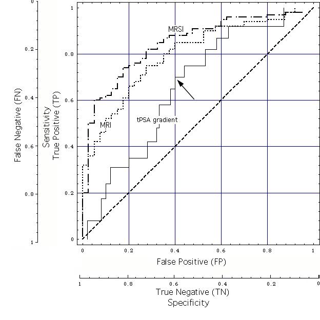
The MRI and MRSI ROCs of Fig. ROC4 are shown here for comparison. Note that -unlike for the tPSA time gradient model- the reference standard for the MRI/MRSI models are whole-mount step-section pathology maps.